Category: General Abstract
1048 - SH2-Flow: A Multiplex Single Cell Phosphotyrosine Profiling Tool for B-cell Malignancies

Introduction: Aberrant activation of tyrosine kinases leads to oncogenic cell signaling, causing cancer cell proliferation, invasion, and metastasis; targeted tyrosine kinase inhibitors of the B cell receptor (BCR) pathway, particularly BTK inhibitors, have shown promising clinical results in chronic lymphocytic leukemia (CLL) and non-Hodgkin's lymphoma. However, resistance to BTK inhibitors, possibly due to de novo mutations in BCR signaling molecules, and relapse after long-term therapy are problems in clinical practice. Therefore, it is important to monitor changes in pathogenic signaling pathways before, during, and after chemotherapy throughout disease progression. There are many prognostic and therapeutic markers based on gene mutations and protein expression, some of which have been successfully incorporated into clinical practice. However, BCR signaling markers that directly reflect the activation status of signaling pathways have not yet been established. We previously developed SH2 profiling to detect changes in the global tyrosine phosphorylation state of cancer cells using the SH2 domain, a major pTyr-binding module, as a probe. We hypothesized that the phosphotyrosine phosphorylation profile measured by SH2 profiling is a promising tool for monitoring the BCR pathway in B-cell malignancies. The primary goal of current study is to develop a clinically adoptable single-cell phosphotyrosine-SH2 profiling assay platform integrating advanced flow cytometry technologies. The new assays (termed SH2-Flow) is multiplexed, largely automated, and optimized for detecting SH2–pTyr interactions in B-cell malignancy cells at the single cell level.
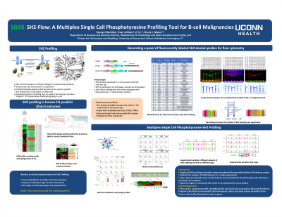
Results: First, we established a flow cytometry-compatible "BCR-SH2 panel" containing 36 SH2 domains involved in BCR signal regulation (Table). The probe protein consists of an N-terminal GST and a C-terminal Halo-His tag, which can be conjugated with a fluorescent dye. Global measurements of protein expression and solubility showed that the majority of proteins are expressed in soluble form. PTyr-binding activity was confirmed by an established quantitative dot-blot assay using stimulated and unstimulated cancer cells. Purified domains were conjugated to fluorescent dyes with various emission spectra by on-column labeling or direct lysate labeling. Labeling efficiency and binding activity of the labeled SH2 domains were assessed by cell staining using SDS gels and fluorescent imagers. Two assay formats were developed to perform a multiplex SH2 flow cytometry assay that can easily detect SH2 binding to cell lines and peripheral blood mononuclear cells (PBMCs) with low background. The 4-plex assay was used for analysis of three time points of CLL PBMC samples (pretreatment, short-term treatment and long-term treatment) showing both probe-specific and sample-specific binding patterns; the 24-plex assay employs a barcoding strategy with CD47 antibodies (Figure 1). Using this method, we analyzed four PBMC samples from four CLL patients with various clinical characteristics which indicates the presence of different SH2 domain binding sites in the CLL cells.
Conclusions: We have established a dye-labeled BCR-SH2 panel that can be used for flow cytometry as well as imaging and other SH2 profiling assays. The 4-plex and 24-plex assay formats have been developed, allowing the versatile application of SH2 profiling in research and diagnosis. Preliminary analyses using CLL PBMC have shown that SH2-Flow has the sensitivity and specificity to select patient subgroups that reflect differences in treatment status and clinical course. Efforts are currently underway to further evaluate the value of SH2-Flow in profiling a CLL cohort, which may lead to the discovery of new biomarkers based on signaling status.
- KM
Kazuya Machida, MD, PhD
Associate Professor
UConn Health
Farmington, CT, United States - EJ
- BM
- JY

